Oral Tissue Biopsy

Early detection saves lives. Painless examination and precise biopsy to diagnose suspicious oral lesions.

What to Expect During and After an Oral Biopsy

Most oral growths and lesions are benign, but any unusual area in the mouth should be evaluated to determine the cause. An oral biopsy allows your specialist to examine a small sample of tissue and determine whether further treatment is needed.

During Your Biopsy

A local anesthetic will be used to numb the area before the procedure. Your specialist will then remove either a small sample of the affected tissue or, when appropriate, the entire lesion. The procedure is typically brief, and the area may be closed with dissolvable sutures.

The tissue sample is sent to a pathologist for careful examination. Once the results are available, our team will discuss the findings with you and coordinate with your general dentist if additional treatment or follow-up is recommended.

After Your Biopsy

Most patients experience only mild discomfort or swelling following an oral biopsy. Any soreness can generally be managed with over-the-counter pain medication as recommended by your doctor and typically improves within a few days. Many patients are able to return to their usual activities shortly after the procedure.

A follow-up appointment may be scheduled to check the healing area and review your biopsy results, generally within several days after the procedure.

Caring for the Biopsy Site

A small amount of bleeding can occur after the procedure. If this happens, gently apply firm pressure to the area with clean gauze or another absorbent material for approximately 10 minutes. If bleeding continues, please contact our office.

While the area heals, avoid biting or putting unnecessary pressure on the treated area. Continue brushing and flossing as usual, but be gentle around the biopsy site and avoid vigorous rinsing. A gentle saltwater rinse may be recommended to help keep the area clean.
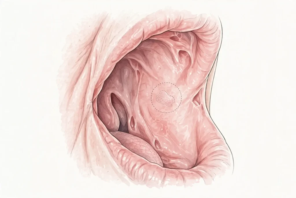
Signs you may need treatment

Red Patches & Sores
White / Gray Lesions

Lumps & Thickening
Abnormal Lesions

What to expect

1

Diagnose

The oral cancer examination is completely painless. Dr. Farhadian will look for abnormalities and feel the face, glands, and neck for unusual bumps. Lasers may be used to highlight pathologic changes and look below the surface for spots and lesions invisible to the naked eye. Over 75% of oral cancers are linked with avoidable behaviors such as smoking, tobacco use, and excessive alcohol consumption.

2

Treat

If abnormalities, lesions, lumps, or leukoplakia are apparent and cannot be diagnosed by history and clinical findings alone, an oral tissue biopsy may be necessary. It is a minimally invasive procedure where a small amount of suspicious tissue is removed, usually under local anesthesia in an outpatient setting on the lip, tongue, palate, or gingiva. Types include incisional biopsy where a small piece is cut from the abnormal area.

3

Restore

Biopsy tissue samples are examined and tested by a pathologist under a microscope for signs of cancer or other issues and precancerous cells. This is the definitive procedure to provide additional information to guide therapy. Based on precise diagnosis, treatment options may include excision, radiation therapy, and chemotherapy. If caught early, oral cancer can be effectively treated.

Common questions

What is an oral biopsy?
A biopsy is a minimally invasive procedure that involves removing a small amount of suspicious tissue from the mouth, usually under local anesthesia, so it can be evaluated closely under a microscope for cancerous or precancerous cells. It is recommended for lesions that last for two weeks or longer after potential irritants are removed.
If during a painless oral cancer exam we find red patches, bleeding sores that fail to heal, leukoplakia (white/gray lesions), soreness, lumps, thickening, or any abnormal lesion, tumor, or mass that cannot be diagnosed by exam alone, we will recommend a biopsy for definitive diagnosis.
The oral cancer exam is completely painless. The biopsy itself is done under local anesthesia in an outpatient setting, so you are comfortable. A small piece of tissue is taken from the lip, tongue, palate, or gingiva and the area heals quickly.
The most traditional is incisional biopsy where a small piece of tissue is cut from the abnormal-looking area. A punch biopsy is a variation. Sometimes a brush biopsy may be used. A wedge-shaped biopsy to capture some normal and abnormal tissue may be preferred to facilitate histopathological examination.
Tissue samples are examined by a pathologist. If caught early, oral cancer can be effectively treated. Based on results, Dr. Farhadian will implement a treatment plan right for you which may include excision, radiation, or chemotherapy, and can provide literature and options for quitting tobacco and other risky behaviors.